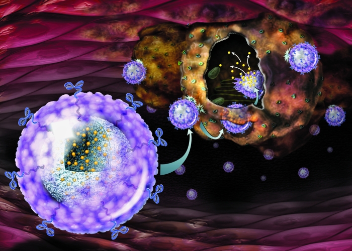
Nanotechnology/nanomaterials for tissue engineering

Hi guys! As we discussed in class this week, tissue engineering aims at developing functional substitutes for damaged tissues and organs. Before transplantation, cells are generally seeded on biomaterial scaffolds that recapitulate the extracellular matrix and provide cells with information that is important for tissue development.

Engineering a functional tissue is difficult. Cells must be organized into tissues with structural and physiological features resembling actual structures in the body. The outer connective tissue that supports cells, known as the extracellular matrix, is especially interesting to us. The matrix and its components — fibers, adhesion proteins, proteoglycans and others — provide cells with a wealth of information that regulates cell growth, shape, migration and differentiation.

To mimic these physiologic features, scientists have been working at the nanoscale – creating structures at the range of 1 billionth of a meter, 100 times smaller than a red blood cell. Through techniques like electrospinning and self-assembly, they can fabricate scaffolds with fibers of the appropriate thickness that form an interconnected, porous architecture. They can modify surfaces by attaching proteins, peptides and other molecules that enhance cell spreading and differentiation. They can increase concentrations of growth factors and cytokines in the engineered tissue by adding charged coatings to which they could stick. Finally, they can modify the 3D shape of the extracellular matrix surface to influence cell shape, differentiation and adhesion.

Nanomaterials can also compensate for limitations in the scaffold. Embedding nanoparticles in biomaterials can enhance their mechanical and electrical conductive properties, for example. Nanomaterials can also increase cell viability, promote adhesion, control the release of growth factors and cytokines, and even physically shape biomaterials and cells to create a desired tissue structure.

Once we engineer a tissue, nanodevices can be useful for triggering desired biological functions and for monitoring engineered tissues before transplantation, to ensure they will function as intended. Nanowires can record the electronic signals that are transmitted through brain cells and cardiac muscle cells for example, or monitor, in real time, the concentration of chemicals of interest within the scaffold. For instance, silicon nanowires with antibodies coupled to their surface have been used to selectively detect biomolecules in serum at femtomolar-level concentrations (a billionth of a millionth of a mole).

Although nanotechnologies have clearly had an impact on tissue engineering, challenges remain. Creating scaffolds with complicated 3D nanoscale surface structures is one challenge. We also need to further explore the biocompatibility and biodegradation of inorganic nanomaterials before they can be safely used clinically.

We’re excited about a future where intelligent nanosensors could be incorporated into engineered tissue to monitor tissue behavior and to trigger the release of biomolecules that promote tissue development and growth. Where nanowired neuroprostheses could assist paralyzed patients by re-routing movement-related signals around injured parts of the nervous system. Where nanomaterials on a tissue’s outer surface could reduce inflammatory responses to transplantation.

Ultimately, smart controllable nanorobots could potentially go to work for us — circulating inside the body, finding diseased tissues and repairing them by destroying defected cells and molecules or by encouraging cells to regain their function. We believe that these tiny nanostructures could redefine medicine in the future. It’s a future I look forward to being a part of.

References:
Dvir, Tal, et al. “Nanotechnological strategies for engineering complex tissues.” Nature nanotechnology 6.1 (2011): 13.
Sethuraman, Swaminathan, Uma Maheswari Krishnan, and Anuradha Subramanian, eds. Biomaterials and Nanotechnology for Tissue Engineering. CRC Press, 2016.

Metallic Scaffolds for Tissue Engineering

Hi guys! This week in class, we learned that the emerging field of tissue Engineering involves replacing, repairing or enhancing biological function at the scale of a tissue or organ by manipulating cells via their extracellular environment.From my understanding, tissue engineering intends to help the body to produce a material that resembles as much as possible the body’s own native tissue.

Scaffolds are often used in tissue engineering. They serve as temporary or permanent artificial Extracellular Matrices (ECM) to accommodate cells and support 3D tissue regeneration. They can also serve as delivery vehicles for exogenous cells, growth factors and genes and as a matrix for cell adhesion. They structurally reinforce the defect to maintain the shape of the defect and prevent distortion of surrounding tissues and act as a barrier to prevent the infiltration of surrounding tissues that may impede the regeneration process.

In general, the requirements of scaffolds for tissue engineering are:
(i) Three-dimensional and highly porous with an interconnected pore network for cell growth and flow transport of nutrients and metabolic waste.
(ii) Biocompatible and bioresorbable with a controllable degradation and resorption rate to match cell/tissue growth in vitro and/or in vivo.
(iii) Suitable surface chemistry for cell attachment, proliferation, and differentation .
(iv) Mechanical properties to match those of the tissues at the site of implantation.


Figure 1. Examples of trabecular scaffolds

Metallic scaffolds have been widely used in tissue engineering because of their good mechanical properties. The main disadvantage of metallic biomaterials is their lack of biological recognition on the material surface. To overcome this restraint, surface coating or surface modification presents a way to preserve the mechanical properties of established biocompatible metals improving the surface biocompatibility. Another limitation of the current metallic biomaterials is the possible release of toxic metallic ions and/or particles through corrosion or wear that lead to inflammatory cascades and allergic reactions, which reduce the biocompatibility and cause tissue loss.

One example of metallic scaffold is tantalum scaffold. Porous tantalum is a biomaterial with a unique set of physical and mechanical properties.It has a high-volume porosity (>80%) with fully interconnected pores to allow secure and rapid bone ingrowth. Studies have demonstrated substantial cortical bone ingrowth between the trabecular network as well as high levels of bone growth onto the scaffold itself. Initial stability of the trabecular metal itself is also higher than that of standard materials, such as cobalt chrome. Furthermore, this new material offers better osteoconduction than other technologies used for biological fixation.


Figure 2. Tantalum scaffolds

Titanium is found to be well tolerated and nearly an inert material in the human body environment.In an optimal situation titanium is capable of osseointegration with bone. In addition, titanium forms a very stable passive layer of TiO2 on its surface and provides superior biocompatibility. The nature of the oxide film that protects the metal substrate from corrosion is of particular importance. In general, porous titanium and titanium alloys exhibit good biocompatibility. Bioactive titanium meshes have been successfully used in spine fusion surgery for the past two decades.


Figure 3. Titanium scaffolds

Future directions of research in metallic scaffold will probably focus on the efficient combinations of osteoinductive materials, osteoinductive growth factors and cell-based tissue regeneration approach using composite constructs carriers to reconstruct and repair hard tissues. The goal is to obtain a functional replacement of the injured hard tissue in a procedure that avoids the step of bone harvesting.

References:
[1] Meyer, Ulrich, et al., eds. Fundamentals of tissue engineering and regenerative medicine. Springer Science & Business Media, 2009.
[2] Pallua, Norbert, and Christoph V. Suschek, eds. Tissue engineering: from Lab to Clinic. Springer Science & Business Media, 2010.
[3] Alvarez, Kelly, and Hideo Nakajima. “Metallic scaffolds for bone regeneration.” Materials 2.3 (2009): 790-832.

Bulk Metallic Glasses for Biomedical Applications

Hi Class! Last week, Ayomiposi Loye gave us a talk on Bulk Metallic Glasses (BMGs) for Biomedical Applications. I feel really excited to learn that BMGs have so many applications in biomedical devices. This is mainly because BMGs have good mechanical properties and biocompatibility, and the ability to be fabricated into desired shapes, which are ideal for versatile implant applications.

Unlike most metals, BMGs have a tendency to avoid crystallization when solidified. It is their amorphous structure that yields many advantages including remarkable properties of high strength (three times that of steel), elasticity, corrosion resistance and durability – all of which exceed that of currently used biomaterials. Most notable, however, is their unique processability that allows them to be molded like plastics with nano-scale precision and complex geometries.

Related image

This processing capability has only come with the recent emergence of thermoplastic forming (TPF), which decouples the fast cooling process from the molding process, allowing the time needed for precision net-shaping. For biomedical applications, biocompatibility of BMGs must also be considered. Their in vitro and in vivo study results indicated that the BMGs are compatible with cell growth and tissue function. Moreover, the ability to independently vary the chemistry, atomic structure, and surface topography – unique to BMGs – revealed the individual contribution of each on biocompatibility.

Researchers have targeted three applications: (1) bone replacement, (2) soft tissue implants (i.e., stents), and (3) surface patterning to program cellular response (i.e., synthetic membranes such as artificial kidneys). It is important that the shape and bulk properties of biomaterials mimic the tissues they replace and that surface chemistry and topography elicit the appropriate cellular response.

In a nutshell, BMGs are a promising biomaterial due to their superior mechanical properties and corrosion and wear resistance over currently metallic biomaterials. The in vitro and in vivo results indicate that the BMGs are in general nontoxic to cells and compatible with cell growth and tissue function. TPF-based processing methods for BMG were developed which satisfy the required precision and repeatability to shape intricate geometries used in biomedical applications. The ability to precisely net-shape complex geometries combined in a single processing step with patterning the surface will enable us to program desirable and predictable cellular response into a 3-D biomaterial.

References:
[1] Schroers, Jan, et al. “Bulk metallic glasses for biomedical applications.” JOM 61.9 (2009): 21-29.
[2] Li, H. F., and Y. F. Zheng. “Recent advances in bulk metallic glasses for biomedical applications.” Acta biomaterialia 36 (2016): 1-20.
[3] Greer, A. L., and E. Ma. “Bulk metallic glasses: at the cutting edge of metals research.” MRS bulletin 32.8 (2007): 611-619.

Metallic Glass – glassy structures of metallic components

Hi Class! This week, we talked about crystal structures versus glassy structures. For crystals, we know there have to be nucleus forming followed by diffusion and subsequent growth to form very ordered atomic structures. And Prof. Gonzalez mentioned if we have a highly viscous liquid, complex crystal structure, and cooled rapidly, formation of glasses will be favorable. After class, I learned that normally crystalline metals can also form glassy structures if they are cooled down very rapidly. I am gonna introduce in this week’s blog this new kind of materials, metallic glasses.

In the past, small batches of amorphous metals with high surface area configurations (ribbons, wires, films, etc.) have been produced through the implementation of extremely rapid rates of cooling. This was initially termed “splat cooling” by doctoral student W. Klement at Caltech, who showed that cooling rates on the order of millions of degrees per second is sufficient to impede the formation of crystals, and the metallic atoms become “locked into” a glassy state. Amorphous metal wires have been produced by sputtering molten metal onto a spinning metal disk. More recently a number of alloys have been produced in layers with thickness exceeding 1 millimeter. These are known as bulk metallic glasses (BMG).  Figure 1 shows the TTT diagram of a metallic glass. Crystallization rate is determined by the competing effects of undercooling and reaction kinetics. Note supercooled liquid region exists between the melting and glass transition temperatures over time periods not exceeding that required for crystal nucleation.

Related image

Figure 1.Schematic time temperature transformation (TTT) diagram for metallic glasses.

Lacking the dislocations and grain boundaries inherent in crystalline materials, metallic glasses exhibit physical properties representative of a completely new paradigm in materials science. Without the premature deformation of slip, elastic strain may regularly approach 2% thereby facilitating strength and hardness values which are far beyond those of crystalline metals. (Figure 2) According to literatures, the Young’s Modulus of metallic glasses can exceed that of metals. Further to such desirable mechanical properties, metallic glasses exhibit a full range of toughness values, low mechanical damping, good corrosion resistance and high magnetic permeability coupled with low coercivity to give superior soft magnetic properties.

page3image717947008
Figure 2. Schematic representation of room temperature yield (metals, composites and polymers) or flexural strength (ceramics) as a function of modulus

In a nutshell, as a class of materials, amorphous metallic materials uniquely
combine high strength and resilience coupled with improved corrosion resistance and the ability to be superplastically formed at relatively low temperature.

References:
  1. Klement, Jr., W.; Willens, R. H.; Duwez, Pol (1960). “Non-crystalline Structure in Solidified Gold-Silicon Alloys”. Nature. 187(4740): 869. 
  2. Liebermann, H.; Graham, C. (1976). “Production of Amorphous Alloy Ribbons and Effects of Apparatus Parameters on Ribbon Dimensions”. IEEE Transactions on Magnetics. 12(6): 921. 
  3. Burgess, Tim, and Michael Ferry. “Nanoindentation of metallic glasses.” Materials Today12.1-2 (2009): 24-32.

Cell interactions with nano-patterned metallic implants

Hi Class! This week, we learned the interaction of nano-or micro-scale biomaterials with cells and the interaction of cells/tissues/organism with large scale biomaterials at both the cellular level and the tissue/host level. In this blog, I am going to talk about cell interactions with nano-patterned implants with a focus on implants made of metallic materials.

Metallic materials are often used for fabricating orthopedic and dental implants because their surfaces are biocompatible with tissues at the target area. Some commonly used surface modification methods for metals include grid-blasting, acid etching, chemical grafting, ionic implantation and calcium phosphate coatings. These strategies for modifying the nature of this interface usually involve changes to the surface at the nanometer level, thereby affecting protein adsorption, cell–substrate interactions, and tissue development.

page13image1232813920

Figure 1: Tissue – dental implant interactions at both gingival and bone sites.

For example, anodization of titanium implants can yield features of nanometer size dimensions . This is achieved by developing a titanium oxide layer using a platinum counter electrode in acidic solutions at a potential of 5-25 V. Through changing experimental conditions (i.e. potential, temperature, electrolyte), scientists have produced an oxide layer a few microns in thickness and composed of a regular array of nanometer sized pores with diameters in the 30-100 nm range, growing perpendicular to the titanium surface. The pore sizes are compatible with those of proteins such as fibronectin (FN) and vitronectin which play an important role in cell adhesion, as discussed in class.

In a nutshell, careful preparation of standardized nanostructured surfaces with repetitive topography may elicit protein adsorption, cell response and cell differentiation. Further research aiming at correlating cell behavior and tissue integration in the future will help us understand the role of surface nanostructures in these biological responses. It is possible to imagine controlling peri-implant tissue healing by changing the surface properties at the nanometer scale.

References:

[1] Lavenus, Sandrine, et al. “Cell interaction with nanopatterned surface of implants.” Nanomedicine 5.6 (2010): 937-947.

[2] Ratner, Buddy D., et al. Biomaterials science: an introduction to materials in medicine. Elsevier, 2004.

Metallic nanoparticles for drug delivery applications

Hi Class! This week, Prof. Zhou talked about the importance of drug delivery in medical sciences and biomaterials used for drug delivery with a focus on polymers and polymeric nanoparticles. This blog post will look at another excellent material candidate for drug delivery, which is metallic nanoparticle/nano-device.

First, here is a brief introduction of the topic as well as a recap from the lectures. Drug delivery is one of the most important research directions in biomedical sciences. The increase of the drug efficacy and the reduction of the side effects are among the goals of the research in the field. Researchers have been working on the development of metallic nanoparticles which target the infected cells without doing harm to other neighbor cells. Metallic nanoparticles such as gold nanoparticles, silver nanoparticles, iron nanoparticles and copper nanoparticles are being extensively researched which show high potential in site-specific drug delivery. These nanoparticles have unique physical and chemical properties such as, plasmatic resonance, fluorescent enhancement, and catalytic activity enhancement which make them very fascinating materials in drug delivery applications. [1]

Figure 1. A schematic of gold nanoparticles for drug delivery to cells [1]

From the lectures, we learned that the performance of nanoparticles inside the human body is controlled by complex factors such as their shape, density, size distribution and surface characteristics. All these factors control the flow properties of nanoparticles, bifurcation in the vascular compartment, modulation of circulation time, and mode of entry into the cell.

Take gold nanoparticles as an example, they can bind with a wide range of organic molecules due to their surface properties so they have been used as therapeutic agents or vaccine carriers into the specific cells. Gold nanoparticles having small size and diameter about 1 nm are able to cross the cell membrane and nucleus to interact with DNA. Therefore by using gene gun, gold nanoparticles have been extensively used for epidermal delivery of DNA vaccines and this method is one of the best methods to deliver DNA vaccine. Another application is that coated walls of gold nano-cages with temperature-sensitive polymer were used as drug carrier which releases their effectors with interaction of near-infrared irradiations. [2]

Image result for gold nanoparticle

Figure 2. A schematic of gold nanoparticles for different applications [3]

Compared to traditional drug delivery methods, some advantages of gold nanoparticles are summarized as follows :

  1. Gold nanoparticles have unique optical, physical and chemical properties due to their size and shape
  2. Gold nanoparticles have high surface area which provide dense drug loading;
  3. These particles are biocompatible and are readily available for conjugation with small biomolecules such as proteins, enzymes, carboxylic acid, DNA, and amino acids;
  4. Due to small size and uniform dispersion they can easily reach to the targeted site with blood flow;
  5. They are non-cytotoxic to the normal cells;
  6. Gold nanoparticles are easily synthesized by various methods.

As pointed out in a review article [4], recent advances in the field of metallic nanoparticles offer the promise of better diagnostic and therapeutic options. Metallic nanoparticles are attracting attention in drug delivery because of their superb properties for targeted delivery in imaging and drug delivery to the desired site. We will continue to see breakthroughs in research and technology of drug delivery based on metallic nanotechnology in the future and these fascinating nanoparticles will indeed provide a new approach to effective and precise drug delivery.


References:

  • [1] Website: http://nanoparticles.org
  • [2] Khan, A. K., et al. “Gold nanoparticles: synthesis and applications in drug delivery.” Tropical Journal of Pharmaceutical Research 13.7 (2014): 1169-1177.
  • [3] Hussain, Kashif, and Touseef Hussain. “Gold nanoparticles: a boon to drug delivery system.” South Indian Journal of Biological Sciences 1.3 (2015): 128-133.
  • [4] Ahmad, Mohammad Zaki, et al. “Metallic nanoparticles: technology overview & drug delivery applications in oncology.” Expert opinion on drug delivery 7.8 (2010): 927-942.


Topic of Interest: Metallic Biomaterials

Hi Class! I am interested in metallic materials for biological and medical uses. Metallic biomaterials can be metals, alloys, metallic glasses, and other forms of materials. They have been widely studied and used due to their excellent thermal conductivity and mechanical properties. Metallic biomaterials have broad applications (see Figure 1). For example, they are used as structural materials for medical devices in the fields of orthopedic surgery, blood circulatory system, and dentistry.

During this semester, I will investigate some of the commonly used metallic biomaterials and also keep an eye for the latest research updates on this topic. I will explore the properties of metallic biomaterials such as mechanical properties, surface/interface properties, and biocompatibility. Applications of this class of biomaterials in the fields of medicine, dentistry, and biotechnology will also be surveyed and introduced. Besides, I am also interested in comparing metallic biomaterials with other popular candidates for biomedical uses including polymers, ceramics, etc.

Image result for metallic biomaterials

Figure 1. Device applications of metallic biomaterials