The Possibilities of Tissue Engineering for Bone Regeneration Using Bioactive Biomaterials

Blog 4: Week of 10/21/2018-10/27/2018: for this week, using lecture ten, I will discuss how can we potentially employ tissue engineering for bone regeneration using the bioactive biomaterials discussed. 

The endoskeletal system is an essential system that facilitates movement,  load-bearing role, and providing the structural maintenance and for the protection our most vital and delicate organs such as the brain and heart.

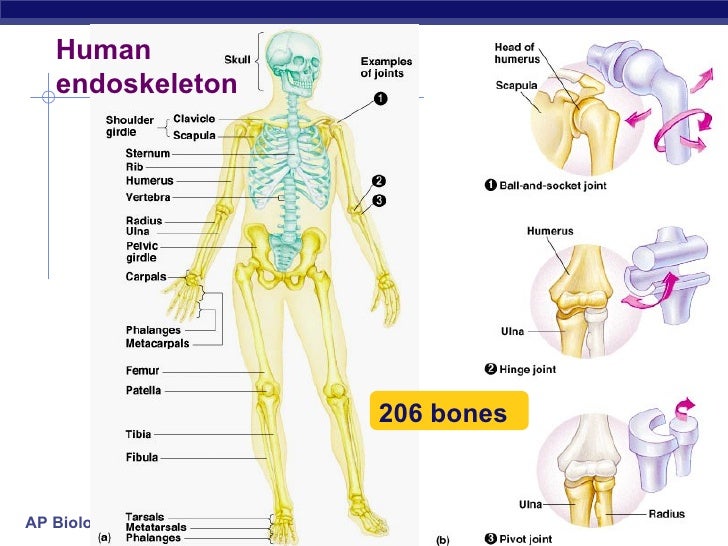
Figure 1: the human endoskeleton system consisted of 206 bone that provides structural support, organ protection, ion homeostasis, and site of immunological cell regeneration and maturation.

In addition to these primary structural supports, it serves a key function in Ca and P homeostasis, and function as the site of immunological cells generation (the bone marrow for hematocrit regeneration). The homeostasis of Ca and P play a significant role in signal activation or inactivation throughout the various muscle types in the bone. Consequently having a misfunction of a bone tissue induces huge complications in one life..causing one to lose one or more of the above inherent life functions of the bone tissue.

In order to alleviate these issues, biomaterials have been employed in different hallmarks.  Nevertheless, the proposed implantation of these biomaterials in the past mostly has been in the form of a bio-inert material(1). And so, the contemporary consideration biomaterials to incorporate into tissue engineering methods, toward a design that employs bioactive materials that will integrate with biological cells for the regeneration of the bone tissue. Before addressing how tissue engineering could be employed for bone tissue regeneration, it’s relevant to discuss the fundamental organizations regarding the bone tissue.

The bone is made of bone matrix and the cells that made it. The bone matrix consists of osteoid, an organic material which includes various proteins and mainly collagen type I which dictates the tensile strength of the bone. The second constituent of bone matrix is hydroxyapatite, an inorganic mineral which comprises calcium phosphate crystals which dictates the rigidity and density of bones.

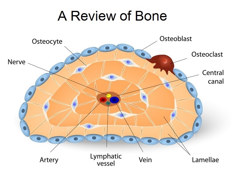
Figure 2: bone cell types that synthesis the bone matrix which serves as the framework for bone regeneration.

The cells that made matrix can be grouped into three main groups. The osteoprogenitor cells which are the precursor for the osteoblast cells which growth factors. The first types of cell that build the matrix are the osteoblast cells, which builds (synthesis) the two matrix components which are the primary structures for bone tissue regenerations.  These osteoblast cells then matured towards osteocytes which occupy space in the matrix with sensors for bone cellular communication (are considered mechanosensory cells of the bone). And the last types of bone cells are osteoclasts that crash bones matrix to release Ca and P ions which play in reabsorption of bone.

The osteoblasts build a bone while the osteoclasts crash the bone. Thus the bone is under a constant remodeling homeostasis. The implication of the remodeling of the bone is that it keeps the homeostasis of Ca and P ions in the bone. When calcium is needed in the body for signal activation or inhibition, the osteoclasts crashes bone to release these ions and when the body has high Ca and P ions, the osteoblast take up these ions to build the bone.

So, knowing the physiological regeneration of bone tissue, how can we use biomaterials to regenerate bone tissues in order to substitute degraded bones? Given the high demand of clinical need of biomaterials for orthopedic, we  can also tailor the biomaterials to be osteoinductive (able to support the differential of progenitor cells to an osteoblast lineage),  osteoconductive (cable to promote bone growth and encourage the ingrowth encompassing bone) and finally osseointegration (cable of integrate within the surrounding bone) (2).

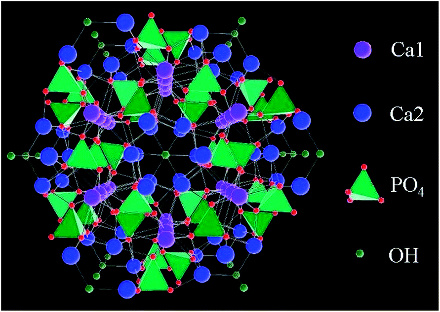
Figure 3: Hydroxyapatite, the major consist of the bone tissue matrix and a major candiate of biomaterirals in bone tissue engineering.

Therefore,  biomaterials are required to be bioactive materials (including bioactive ceramics, bioactive glasses, and or a combination with polymers) to be incorporated in bone tissue engineering. Using these bioactive materials, the scaffold (framework) will be built in a way that over time, these materials will be biodegraded slowly and replaced with the patient’s own newly regenerated bone tissues.

Generally speaking, the bioactive inorganic materials that encompass tricalcium phosphate, bioglasses, and their combination with polymers can be tailored to design varies characteristics of a specific scaffold. Once scaffold is designed with these bioactive and biodegradable materials, various growth factors will be delivered to the scaffold using nanoparticle in order to enhance the regeneration of bone tissues as shown below (3).

Figure 4: different mechanism of introducing growth factors in the bone matrix to facilitate bone tissue regeneration

Finally, in modeling the bone tissue engineering, if we can master the modeling of biological complexity with three-dimensionally synthetics bioactive materials and the and closely mimic the physiological communication complexity of cells in the matrix, we can potentially expedite the field of bone tissue engineering to regenerate patient own bone tissue that will solve the incompatibility issue including immunological  reactions due to the introduction of bio-incompatible materials in the body.


Sources:

  1. Lanza, Robert, Robert Langer, and Joseph P. Vacanti, eds. Principles of tissue engineering. Academic press, 2011.
  2. Stevens, Molly M. “Biomaterials for bone tissue engineering.” Materials today 11.5 (2008): 18-25.
  3. Lee, Soo-Hong, and Heungsoo Shin. “Matrices and scaffolds for delivery of bioactive molecules in bone and cartilage tissue engineering.” Advanced drug delivery reviews 59.4-5 (2007): 339-359.

Is Re-engineering the Activation and Deactivation of the Immunity System for Control Cell Death or Cell Regeneration Possible?

Week of 10/1/2018-10/06/2018

Engineering cells for the regeneration of a damaged tissue such as blood vessel or for the eradication of diseases causing agents in tissues such as cancer are central themes in tissue engineering. For this week, using lecture eight, I will discuss how can we potentially re-engineer macrophage activation pathway so that we can specifically deactivate this pathway to regenerate a damaged tissue or activate to cause cell apoptosis in cancer cells.
In order to manipulate the cellular machine inside a cell, we have to transmit a chemical molecule that would alter the mechanism of the cellular machine. To engineer a particular tissue to regenerate, a signal has to be sent to alternate the activation of macrophages for wound repair, tissue fibrosis because activation of macrophages causes the activation of adaptive immune systems such as T-cells and B-cells which leads cell apoptosis in a downstream activation. There are substantial challenges in the way of getting a  drug to target organelle and if we can somehow overcome these barriers we can potentially achieve the desired effects in a cellular and tissue level.

The first issue is that the undesired cellular uptake: that’s  when a drug is circulated in a circulatory system, the broken up peptides on the surface of our engineered nanoparticles that carries our drug will be detected by the immunity system and will be sent for degradation to different parts of the organ depending on the location of detection and the type protein coated on the surface of the nanoparticle. We can overcome this barrier by implementing various strategies.

Figure 1: PEGylation of nanoparticle for the improvement of blood circulation time

One of the strategies to use the mechanism of PEGylation, a process of attaching a PEG molecule covalently or non-covalently onto a nanoparticle to reduce the interaction of the antigenic protein on the surface of the nanoparticle with the immune system cells (1). This molecule reduces the number of protein on the surface of the nanoparticle thus reducing the cellular uptake of the nanoparticle with the drug as shown in figure 1. This mechanism merely opened the gateway of solving  the cellular uptake problem,  and since then,  a more robust and improved version of this mechanism has been developed: one of  which is to use a self-peptide thus the cell will recognize that peptide as  its peptide so that it doesn’t trigger the immune system and thus substantially reduces the cellular uptake of that molecule.

However, the use of self-peptide only solves just one of the drug delivery issue. Another issue is that we want cellular uptake of our drug but still we want to deliver our drug to a target cell and a target organelle in that cell. That’s we have to have a system to overcome the cellular semi-permeability layer barrier because the membrane is semipermeable to the selected molecule: it doesn’t just let in any molecule into the cells. It acts as one of the primary mechanism to protect the cell and regulate the movement of the molecule into and out of the cell using a sophisticated membrane protein integration.

Figure 2: The HIV TAT penetrates the cellular membrane without inducing a cellular damage

 To solve this problem, we need to acquire understanding how viruses such HIV gets into the cell.  The HIV is one of the sophisticated molecule machinery that hijack the human cellular regulations to potentially get into the cell and use the human cellular machinery to make its own necessary protein and other molecules by inserting its DNA into the human DNA. In early 1990,  researchers were able to recognize a sequence of a protein that the virus employed to enter into the cell as shown in figure 2: cell penetrating peptide (CPP) called HIV TAT sequence (2). That is another incredibly amazing achievement by the virus. When I studied about this mechanism, I always amazed how the sophisticated cellular machinery regulation by using proteins and molecular chemistry help sustain life this long against hypermutation cable virus molecule. As the size of a cell or a molecule (such as human cell compare to single bacteria compare to the virus), the speed of evolution increase.  The virus is just a molecule which is considerably small compared to a human cell, thus having the enormous advantage of hypermutation in a short period of time to hijack the cellular regulations. It seems to me that without the human substantial effort in research to the breakthrough in understanding the cellular mechanisms, the speculation of cell surviving in the competition of virus would be difficult just like the HIV epidemic would have killed most of the human species. Thank you for the scientific research and discoveries, now we know a lot about cellar mechanism even though we have to go a lot to specifically modulate the cellular regulations in tissue engineering. Now let’s get back to the idea of learning from the virus, now we can hijack the virus mechanism for our advantage in tissue engineering. Using the peptide sequence that the virus uses and with even a more improved version, we can now send nanoparticle with the drug into the cell without inducing damaging to that cellular membrane.

At this point, the first two major barriers are solved. The next issue is that once the nanoparticle gets into the cell, the cell immediately captures the nanoparticle with liposome for vesicle (pack) and to send it to lysosome to degrade the molecule. Again this regulation has a substantial evolutionary advantage because the cell uses this mechanism to clean up detrimental material in the cell because the cell doesn’t have a recognition what that nanoparticle contains (the nanoparticle gets into the cell without any regulation of the cell and so the nanoparticle has to deal with the fate of degradation).

Figure 3: endosomal escape mechanism to deliver a drug to a target organelle in the cell.

For this barrier, a lot of agents have developed with some side effect and benefits to altering the liposome function before the nanoparticle gets to the lysosome for degradation.  One of the mechanisms we can use for this purpose is the endosomal escape through membrane fusion to release a cargo containing the drug for a target molecule in a cell as shown in figure 3 (3).


Finally, once we passed the last stage, we can engineer our nanoparticle to go into the target organelles such as mitochondria, nucleus…or to exit the cell. For our purpose, at this stage, we can deliver a drug to perform the two ultimate goals. The first goal is to increase tissue regeneration for wound repair via tissue fibrosis by deactivating the macrophage presentation (protosome degradation and presentation). Or the second ultimate goal which is to specifically activate the macrophages in the cancer cell to increase cellular inflammation via T-cells and antibodies to potentially cause cancer cells apoptosis.

Source:

  1. Suh, Junghae, et al. “PEGylation of nanoparticles improves their cytoplasmic transport.” International journal of nanomedicine 2.4 (2007): 735.
  2. CPP Peptide Synthesis: Make Your Peptide Cell Permeable!www.lifetein.com/Cell_Permeable_Peptides.html.
  3. Lönn, Peter, et al. “Enhancing endosomal escape for intracellular delivery of macromolecular biologic therapeutics.” Scientific reports 6 (2016): 32301.