Degradable Natural Polymers

Stitches can be required following contact in various sporting events. This occurs at every level from the professional to the  recreational. In a 2016 basketball contest for the Miami Heat of the NBA, Goran Dragic left the game bloody, as seen in figure 1, after taking an elbow to the face. He returned to the floor in the second half with 8 stitches in his lip. An example that shows this occurring in a more “casual” setting is illustrated in figure 2 which depicts that in 2010 Barack Obama also caught an elbow and required 12 stitches to the lip. A desirable material to perform these stitches would be strong enough to hold tissue together and last long enough to allow the body to heal itself. For the examples provided it would also be beneficial if the patient didn’t have to go to the doctors again to get the stitches removed. Therefore natural polymers are a good choice for this application because they can be disposed of by normal metabolic processes.

Figure 1. “Heat guard Goran Dragic leaves the floor for treatment of a cut lip after being called for a foul against Toronto Raptors in Game 2” of 2016 Eastern Conference semifinal. (Dan Hamilton/USA Today Sports)

Figure 2. President Barack Obama walks to his car after a game of basketball in 2010 where he suffered a busted lip. (Tim Sloan/AFP/Getty Images)

In a 2011 paper, Catechol-Functionalized Chitosan/Pluronic Hydrogels for Tissue Adhesives and Hemostatic Materials, Ryu et al. showed that the natural material chitosan could be used to create a hydrogel that could be used as a tissue adhesive. Figure 3 shows a diagram of how this hydrogel would work and figure 4 demonstrates its adhesion properties and how they were tested.

Figure 3. Schematic representation of tissue adhesive, thermosensitive, and in situ cross-linkable CHI-C/Plu-SH hydrogels. (Ryu et al. Biomacromolecules. 2011)

Figure 4. In vitro quantitative adhesion analysis of hydrogels. (A) Illustration of tissue adhesion test using a universal testing machine. (B) Detachment stress of various compositions (Ryu et al. Biomacromolecules. 2011)

Al-Mubarak et al. state in their 2013 paper, Cutaneous Wound Closure Materials: An Overview and Update, that”there are many advantages of tissue adhesives over suturing and other methods of wound closure, such as a lower infection rate, less operating room time, good cosmetic results, lower costs, ease of use, immediate wound sealing, faster return to athletic and work activities, elimination of needle-stick injuries and eliminating the need for post-operative suture removal.” With these benefits in mind, tissue adhesives  made of natural materials may become the new and preferred way to treat lacerations that occur from sports.

Carbon Fiber Prosthetic

The integration of carbon fiber reinforced polymer (CFRP) composites into the structure of prosthetics has changed the way that amputees are able to participate in sports.Figure 1shows the first below-knee prosthesis made purely from CFRP, the Flex Foot, which was  developed by Phillips in 1985. The mechanical properties of the composite including its lightweight, flexibility and high strength are what make it a good material to use for this application. Those properties allow for energy to be stored when the compressive load of a person’s weight is put on the prosthetic. Lifting body weight off as in a step results in decompression and the material returns to its original shape releasing the stored energy. So the carbon fiber composite stores elastic energy while in the stance phase and releases it during the step which aids in the swinging of the leg. This energy store and release system is what enhances a prosthetic’s performance.  When describing  how the amputee/prosthetic system behaves one could assume that it behaves like a perfect spring meaning Hooke’s law would apply and there’d be a linear relationship between stress and strain and no energy would be lost. However, a more realistic model would account for sources of energy loss such as friction, heat and sound. 

Figure 1. Schematic representation of the Flex Foot design (Scholz M.S. et al. Composites Science and Technology. 2011)

In terms of the make up of the material, the carbon fibers imbue strength and the polymer contributes its flexibility.  The concept of property averaging explains that the properties of a composite represent some average of the constituent properties. Because fibers respond in a different mechanical manner  when load is applied parallel  to their orientation vs transversely, the average material property is sensitive to geometry. Therefore, not only is the design of the prosthetic important for its function, but in order to tune performance for individuals the materials composition can be changed. Adjustments that can be made for possible customizations include differing laminate thickness and or fiber orientation.

In conclusion, the mechanical properties of CFRP make it a good material to design prosthetics with that can closely match human limb performance. An  example of what this means in terms of sports and competition is that in 2012 Summer Olympics Oscar Pistorius, shown in figure 2, became the first amputee runner to compete at an Olympic game.

Figure 2. Oscar Pistorius in the Men’s 4×400-Meter Relay, Track and Field finals at the 2012 Summer Olympics in London (Chang W. Lee/The New York Times)

Source: M.S. Scholz et al., The use of composite materials in modern orthopedic medicine and prosthetic devices: A review, Composites Science and Technology 2011

Suggesting use of bulk metallic glass as replacement for stainless steel alloys in fracture repair

Based on the literature, corrosion is a problem that leads to implant removal. Therefore I suggest  the use of bulk metallic glasses (BMGs) in orthopedic applications due to their enhanced corrosion resistance and high strength.

Stainless steel is used in medical applications because of how easy it is to form it, its strength, and low cost. This material used in fracture fixation applications is susceptible to pitting and crevice corrosion. According to the 1995 paper Failures in stainless steel orthopaedic implant devices: a survey, “It has been estimated that 10–20% of all stainless steel implants need to be prematurely removed, and while corrosion was identified as the primary cause for nearly a quarter of these premature removals, nearly all instances of implant removal showed evidence of some degree of implant corrosion.”

To show that the body’s response to materials results in corrosion of stainless steel, specifically 316L, Brooks et al. simulated the effects of of inflammation on the materials degradation. Figure 1 shows corrosion because more ions are in solution from the metal in the inflammatory simulation, done by making solution at a low pH with hydrogen peroxide, than in the normal one.

Figure 1. Concentration of ions released from 316L into the test electrolyte over a 24hr immersion period in both normal and inflammatory test conditions. (Brooks et al. Materials Science and Engineering C. 2017)

BMGs therefore could be an alternative for stainless steel because, it is strong, easy to form and corrosion resistant. The various methods of shaping BMGs including blow molding, casting and thermoplastic forming show how they are processable like a polymeric material and at relatively low temperatures. BMGs are super cooled resulting in an amorphous structure. This amorphous structure means that there are no grain boundaries and the absence of them contributes to the material’s corrosion resistance.

Calcium Phosphate Cements in Bone Repair and Regeneration

This post will be a review of a 2017 paper in Bone Research Calcium phosphate cements for bone engineering and their biological properties by Xu et al.

Calcium phosphate compounds are abundant in nature and in living systems and come in many forms.They are all bioactive, osteoconductive, and biodegradable and these properties are what make it a desirable material to be used in bone repair and regeneration applications.

Bioactivity refers to the ability of bone scaffolds to bind directly to the surrounding bone without the formation of fibrous tissue. Since bioactivity is such an important property of calcium phosphate cements (CPCs) , the paper discusses how they have been modified with the addition of bioactive glass.

Osteoconductivity is the property of a material to facilitate the growth of new bone on its surface. The paper discusses the importance of composition in osteconduction and uses an example of where a silicon CPC (Si-CPC) was developed and showed up to threefold osteoblast cell proliferation as compared to the no silicon. Not only does composition effect properties like osteoconduction, but it also plays a role in the solubility of the material which can be important for delivery and mixing.

Additionally, composition changes other properties such as degradation. Because of this, material properties are tunable, and a CPC scaffold should ideally degrade at the same rate that new bone forms. Varying physical factors such as crystal size is another way to tailor degradation.

An example how this material can be applied to sports medicine is in anterior cruciate ligament (ACL) reconstruction. This procedure is done to repair torn ACLs which occur in sports such as football,basketball and soccer due to rapid changes in directions and sudden stops. The surgery involves replacing the injured ligament with a soft tissue tendon graft. The long healing time of these grafts within the the bone tunnels from the procedure is a concern of surgeons. It has been shown however, that due to their resorbability and osteconductivity, CPCs can enhance graft healing within the bone tunnels. Figure 1 illustrates  a schematic for the graft and CPC coating. The image describes an experiment that the group designed in order to investigate how enhancing CPC osteoconductivity with the incorporation of strontium effected graft healing.

Figure 1. “Surgical design. The graft on the left limb was coated with strontium-enriched calcium phosphate cement (Sr-CPC). The graft on the right limb was coated with conventional calcium phosphate cement (CPC). The area of the cement coating on the tendon allograft surface is marked as gray, while the uncoated area of the allograft is marked as white.” (Kuang G.M. et al. The American Journal of Sports Medicine. 2014)


Interface of Academic Research and Commerce Case Study: Football Helmet That Diagnoses Concussions

This post will review the development of a football helmet equipped to diagnose concussions in real time. The work is being done by Drs. Barclay Morrison and James Noble at Columbia University. Specific focus will be placed on the marketability of this project.

Diagnosis of a concussion first requires that symptoms are either noticed and reported by players themselves or others on sidelines. Because players are competitive and feel pressure to perform, they might chose to ignore or dismiss symptoms. As a result,  concussions can be difficult to diagnose when they occur and go unnoticed. Continuing to play after experiencing a concussion puts players at further risk of injury. The researchers at Columbia noticed this problem and posed a solution, a way of diagnosing concussions objectively and in real time by utilizing ECG technology imbedded into helmets.

The basic concept is that ECG electrodes and leads are outfitted into a helmet. The helmet sends a signal to a monitoring device that uses an algorithm that accounts for movement and is able to detect the unique signal produced by a concussion. This allows for clear and informed decision making.  Below, in figure 1 is a flow diagram depicting how the device would be used.

Figure 1. Diagram depicting how ECG technology and data would be used, collected, and analyzed. (NoMo Diagnostics)

Besides meeting the need of better diagnosing concussions, this product has potential to be profitable.  Therefore, it is interesting to examine its development and conception under the lens of patentability.

Helmet development is being funded by the Columbia-Coulter Translational Research Partnership which “provides funding and business support for translational projects that will improve patient care and address unmet healthcare needs”. This program is contributed to by Columbia Technology Ventures which  is analogous to the Office of Cooperative Research at Yale. Morrison and Noble cofounded a company called NoMo Diagnostics and its website reads, the “goal is to develop the first FDA approved, real-time, physiologically relevant concussion diagnostic to initiate early treatment thus avoiding life threatening and long term consequences associated with unrecognized brain trauma.” The source of funding for the project as well as the founding of this company indicate that  this product is marketable.

In order to be patentable an invention must be novel, useful, and non-obvious. The usefulness of a helmet that can diagnose concussions is already addressed above, which leaves the novelty and obviousness to discuss.

If the websites claim is true and this ends up being the first device of its kind, then it is unquestionably novel. The publications about this technology and product do not give many details and the reason for this could be that the company is trying to keep their idea novel in order to be able to patent it. If they were to give out more details, they might provide prior art which would make their patent claim void. An article published by Columbia however, explicitly states that the idea is not completely new  and that “researchers experimented with taping EEG electrodes to the scalps of college athletes” in the 1960’s.

In the case of this product I believe the novelty still remains because the team is taking an existing technology but using it in a new way. Additionally they are finding ways to improve that technology by miniaturizing it and developing new algorithms to analyze the data.

From a biomaterials perspective I’d be interested in knowing what the electrodes and leads are made out of because they would be in contact with human skin in order to pick up a signal. Therefore, their function would have to remain unaffected  by sweat.

Use of polymer nanoparticles to diagnose and treat muscle and tendon injuries

This post will be a review of a 2017 paper in Theranostics, Ultrasonographic Imaging and Anti-inflammatory Therapy of Muscle and Tendon Injuries Using Polymer Nanoparticles by Kim et al.

First off, ultrasonography is a important tool for diagnosing muscle injuries because it allows for the real time visualization of soft tissues at a high resolution. Typically,contrasts agents are used to improve visibility of internal structures. This is done with microbubbles, which have drawbacks because they expand and therefore diffuse at body temperature and are limited to microcirculation and vascularity meaning they have poor tissue permeability. Because of this, work has been done to create particles that generate gas based upon a stimulus, thus producing CO2 by the breaking of carbonate linkages. The group from this paper developed poly(vanillyl alcohol-co-oxalate) (PVAX), a polymer that has this desired reaction . The  CO2 produced by the interaction of the body and PVAX provides image contrasts and the polymer based particles  have a longer half life than other methods.

Additionally, reactive oxygen species such as Hydrogen peroxide (H2O2) are known to be produced by trauma and musculoskeletal injury. PVAX can provide a therapeutic effect to these injuries by reacting with these excess species and releasing bioactive vanillyl alcohol.

This paper then, discusses the potential for a polymer, PVAX, nanoparticle to be used for diagnosis as well as treatment of musculoskeletal injuries which are common in sports.

Properties of PVAX 

PVAX nanoparticles were synthesized using a single emulsion technique.

As can be seen in the chemical structure of PVAX shown in Figure 1, it is a copolymer because the chain is made of different monomers. The molecular weight of PVAX was determined to be ~12,000 Da with polydispersity of 1.6. (Kim et al.) This is important because as discussed in lecture the degradation rate of a polymer depends on its molecular weight. The polydisperity value means that chain lengths have varying molecular mass.


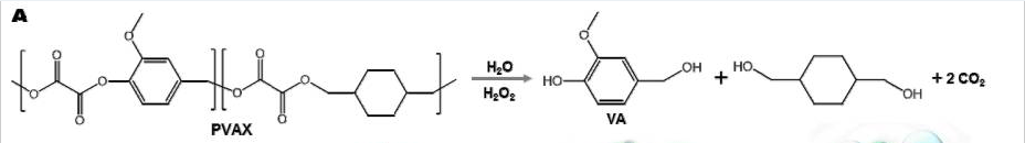
Figure 1. Chemical structure of PVAX and degradation reaction that produces CO2 (Kim G. et al. Theranotics. 2017.)

Biocompatibility means that a material is able to perform its application with an appropriate host response. To test the biocompatibility of PVAX nanoparticles in vitro, cell count was obtained by measuring absorbance before and after a 24 hr incubation. For an in vivo test, the nano particles were injected into mice. The in vitro test did not result in a significant decrease in cell count and the the in vivo test revealed no toxicity to major organs. Taken together, these results show that PVAX nanoparticles have excellent biocompatibility.

Effects of PVAX nanoparticles on ultrasonographic signal

A rat model for contusion  injury was used to investigate how PVAX nanoparticles could improve diagnosis of musculoskeletal injuries.  The experiments determined that PVAX lead to greater contrast in ultrasonographic images with specificity to site of injury where as a current contrast agent SonoVue was non specific. Additionally PVAX nanoparticles increased signal for a longer amount of time than SonoVue.

Therapeutic Effects of PVAX nanoparticles

Reverse transcription polymerase chain reaction was used to investigate the production of pro-inflammatory cytokines such as IL-1β and TNF-αThe data indicating that contusion results in greater expression of these products and the injection of PVAX nanoparticles results in their suppression is seen in figure 2.

Figure 2. Anti-inflammatory activities of PVAX nanoparticles (A) Triceps surae muscles. (B) Achilles tendons. *p<0.05, **p<0.01. (Kim G. et al. Theranotics. 2017.)

Additionally, Western blot assays were used to test for proteins related to cell apoptosis. Contusion lead to up-regulation of Bax and down-regulation of Bcl-2 which is a phenotype associated with apoptosis.  Injection of PVAX nanoparticles suppressed the contusion-mediated apoptosis, evidenced by the up-regulation of pro-caspase-3 and Bcl-2 and down-regulation of Bax. PVAX nanoparticles also reduced production of apoptotic proteins in injured Achilles tendons. These results are significant because Inhibition of caspase mediated apoptosis restores muscle function (Vollmar B. et al. Apoptosis. 2012). The Western plots showing these results can be seen in panels A and C of figure 3.

Figure 3. Anti-apoptotic activities of PVAX nanoparticles. (A) Effects of PVAX nanoparticles on the expression of apoptosis-related proteins in triceps surae muscles. (B) Quantification of apoptosis-related proteins in triceps surae muscles. (C) Effects of PVAX nanoparticles on the expression of apoptosis-related proteins in Achilles tendons. (D) Quantification of apoptosis-related proteins in Achilles tendons. *p<0.05, **p<0.01. (Kim G. et al. Theranotics. 2017.)

Closing Thoughts

This paper gives an example of how the properties of polymers can be used in order to both improve diagnostics and as treatment. The interesting aspect here is that there was no drug imbedded in the polymer, it had the effects purely because of its structure. Something I would try to implement here is changing the shape of the PVAX nanoparticles to see if that had an effect on how it behaves. I hypothesis that changing the surface area would effect the rate of degradation into CO2 thus having an effect on the nano particles use as as contrast agent, but I don’t know how this would effect the therapeutic effects.

The application to sports here is quite useful because an injection of a nanoparticle to improve image quality would begin treatment immediately. An additional benefit, to  improving ultrasonographic imaging, is that you decrease exposure to radiation that typical accompanies other ways of imaging injuries.

Welcome to Biomaterials in Sports!

This blog will explore the use of biomaterials in different sports and why the choice of each material is optimal based on its properties and desired function. I want to pursue this topic because a variety of materials are used to help treat and prevent injuries that arise during participation in sports. The definition of a biomaterial is any substance used for any period of time as a whole or part of a system, which treats, augments or replaces any tissue, organ or function of the body. An example of a material that fits this definition is calcium phosphate cement, used to help heal bone tunneling that occurs during anterior cruciate ligament (ACL) surgery.

Injection of calcium phosphate cement into bone. Image Copyright 2007 by Wright Medical Technology, Inc.